Orthodontics is the specialized branch of dentistry that focuses on diagnosing, preventing, and correcting malocclusions (misalignments) of the teeth and jaws. It is a multifaceted discipline that requires an understanding of both dental and skeletal structures, as well as the interactions between the two. The role of orthodontics extends beyond just cosmetic improvements; it plays a vital role in promoting better oral health, function, and overall quality of life.
Within orthodontics, certain conditions pose more complex challenges, particularly supernumerary teeth (extra teeth) and skeletal malocclusions (abnormal bone growth). These two conditions can disrupt both the alignment of teeth and the development of facial structures. The impact of these conditions can be far-reaching, affecting a person’s ability to speak, chew, and even breathe properly.
Understanding the role of orthodontics in treating these issues involves a detailed look into their causes, symptoms, complications, diagnostic techniques, and treatment options. This comprehensive exploration aims to shed light on how orthodontics can effectively manage these complex dental issues.
Supernumerary Teeth (Extra Teeth)
Supernumerary teeth, also known as extra teeth, are a type of dental anomaly where additional teeth develop beyond the normal 32 teeth in the permanent dentition. They can emerge in any part of the mouth, but are most commonly found in the upper jaw.
A. Classification and Terminology
Supernumerary teeth can be classified based on their location, shape, and the number of teeth involved. The terminology includes several subcategories based on where these teeth develop:
- Mesiodens: The most common type, typically found between the two upper central incisors. They are often small and peg-shaped.
- Paramolars: These are extra teeth that develop next to the molars.
- Distomolars: These teeth appear behind the third molars, resembling additional wisdom teeth.
- Peridens: Located between the primary and permanent teeth, often seen as accessory teeth in the dental arch.
- Supplemental Teeth: Teeth that resemble normal teeth but are additional to the regular set.
B. Causes of Supernumerary Teeth
The exact cause of supernumerary teeth is still not fully understood, though several factors are thought to contribute:
- Genetic Factors: A hereditary predisposition plays a significant role in the development of supernumerary teeth. If a parent or sibling has extra teeth, there is a higher probability that the individual may also develop them.
- Developmental Disruptions: During the development of the teeth, certain genetic or environmental factors may trigger abnormal tooth formation, leading to the growth of extra teeth. These disturbances can be the result of infections, trauma, or hormonal influences.
- Syndromes: Certain genetic syndromes, such as Gardner’s syndrome, Cleft lip and palate, and Crouzon syndrome, have a strong association with the presence of supernumerary teeth. People with these syndromes may experience multiple extra teeth that require special management.
C. Clinical Manifestations and Complications
Supernumerary teeth are not always immediately noticeable, especially if they are hidden under the gum tissue or do not interfere with normal tooth eruption. However, they can lead to significant complications, including:
- Tooth Crowding and Misalignment: Extra teeth can lead to overcrowding by blocking the eruption of adjacent permanent teeth. This results in misaligned teeth that may need orthodontic intervention.
- Delayed Eruption: Supernumerary teeth can prevent the normal eruption of teeth. For instance, a mesiodens may block the path of the upper incisors, leading to delayed or abnormal eruption of the permanent teeth.
- Development of Cysts and Tumors: One of the more serious complications of supernumerary teeth is the potential for the formation of dental cysts or tumors around the abnormal teeth. These cysts, such as dentigerous cysts, can cause bone resorption and damage to adjacent teeth, and may require surgical intervention.
- Infection: If extra teeth cause an obstruction to normal tooth eruption, it may create pockets where food particles and bacteria can accumulate. This can lead to infections and inflammation, further complicating the patient’s oral health.
- Aesthetic and Functional Issues: Supernumerary teeth, especially those that are visible, can affect the appearance of a person’s smile. Additionally, they can interfere with proper chewing and biting, leading to functional problems.
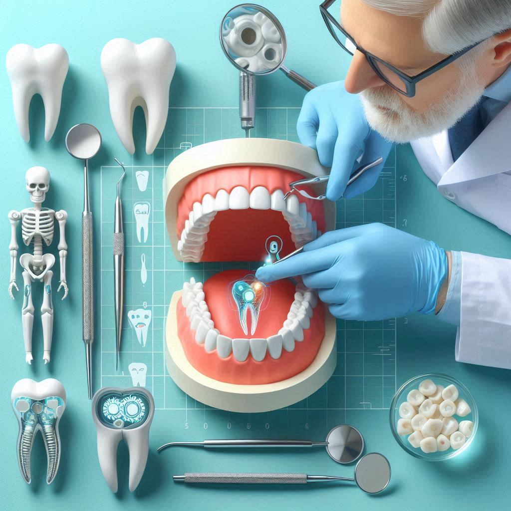
D. Diagnostic Methods
The diagnosis of supernumerary teeth requires careful evaluation using both clinical and radiographic methods. The steps include:
- Clinical Examination: An orthodontist or dentist will visually examine the patient’s mouth to detect any obvious anomalies in tooth eruption or alignment. Palpation (feeling the gums) may also help detect any hidden teeth.
- Radiographs: X-rays, such as panoramic radiographs and intraoral periapical radiographs, provide a detailed view of the location and position of extra teeth. This is essential for identifying impacted teeth or those that are causing developmental disturbances.
- Cone Beam CT (CBCT): This advanced 3D imaging technique is especially useful for locating teeth that are deep within the jaw or obstructing adjacent structures. It provides a detailed, three-dimensional view of the patient’s dental anatomy, allowing for precise treatment planning.
E. Treatment Options for Supernumerary Teeth
Treatment for supernumerary teeth depends on the specific characteristics of the case. Some treatment strategies include:
- Observation: If supernumerary teeth are not causing any immediate issues, the orthodontist may decide to monitor their development over time, especially in young patients who are still growing.
- Extraction: In cases where supernumerary teeth are causing tooth displacement, infections, or blocking the eruption of adjacent teeth, they may need to be removed. Extraction is often the first step in correcting the alignment of the remaining teeth.
- Orthodontic Treatment: Once extra teeth are removed, orthodontic devices such as braces or aligners may be used to reposition the remaining teeth into proper alignment.
- Surgical Intervention: If extra teeth are associated with cysts, tumors, or other complications, surgical removal may be required. This typically involves collaboration between the orthodontist and an oral surgeon.
- Monitoring: For some individuals, especially those with minimal or no symptoms, supernumerary teeth may not require immediate treatment. Regular monitoring with radiographs may be all that is needed until the teeth naturally erupt or any complications arise.
Abnormal Bone Growth and Skeletal Malocclusions
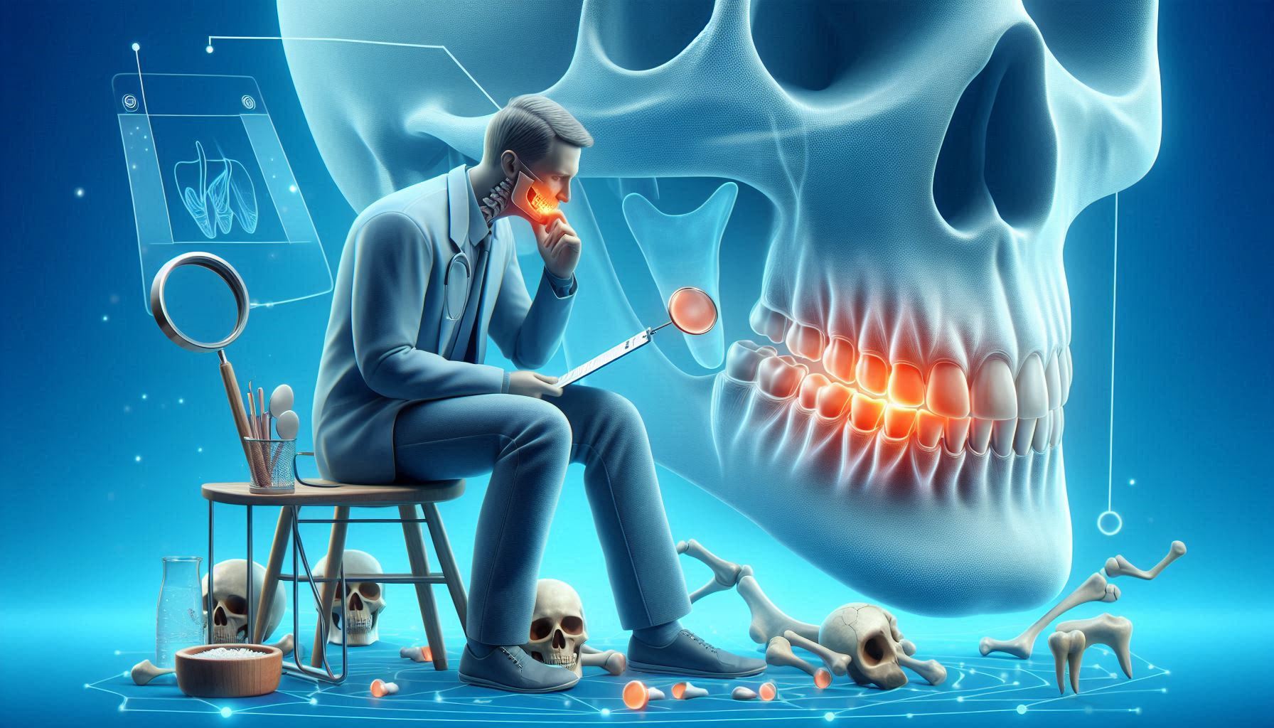
A. Understanding Skeletal Malocclusions
Skeletal malocclusions are irregularities in the alignment of the jaws themselves, as opposed to the teeth. These types of malocclusions can lead to issues with bite, aesthetics, and oral function. Skeletal malocclusions are often classified into three broad categories:
- Class I: Normal jaw relationship with dental misalignment. Teeth may be misaligned, but the jaws are in proper relationship.
- Class II: The upper jaw is too far forward relative to the lower jaw. This condition is also called retrognathism, where the lower jaw appears to be recessed.
- Class III: The lower jaw is too far forward relative to the upper jaw. This condition is known as prognathism, where the lower jaw protrudes significantly.
B. Causes of Skeletal Malocclusions
Several factors contribute to the development of skeletal malocclusions:
- Genetics: Many skeletal malocclusions are inherited. If a parent or sibling has an abnormal jaw relationship, the chances of a child having similar issues increase.
- Developmental Factors: Issues during early jaw growth, such as abnormal positioning of the tongue or thumb sucking, can lead to misalignment of the jawbones.
- Trauma: Accidents or injuries to the face during childhood or adolescence can disrupt normal bone growth and lead to skeletal malocclusion.
- Congenital Conditions: Certain birth defects or syndromes, such as cleft lip and palate or craniofacial disorders, are associated with skeletal malocclusions.
- Environmental Factors: Chronic mouth breathing, thumb sucking, or prolonged pacifier use can affect the development of the jawbones, leading to malocclusions.
C. Types of Skeletal Malocclusions
- Class I Malocclusion: While the jaws are properly aligned, the teeth may be overcrowded, misaligned, or positioned improperly due to factors such as tooth eruption issues or missing teeth.
- Class II Malocclusion: This type is characterized by a receded lower jaw (retrognathism). The upper teeth and jaw are positioned further forward than the lower jaw, leading to an overbite.
- Class III Malocclusion: In this condition, the lower jaw protrudes beyond the upper jaw (prognathism), leading to an underbite.
D. Clinical Effects of Skeletal Malocclusions
The clinical effects of skeletal malocclusions are significant, especially when they interfere with basic functions like speaking, eating, and breathing. Some potential effects include:
- Speech Difficulties: Malocclusions can lead to speech problems such as lisps, difficulty pronouncing certain sounds, and issues with clarity.
- Chewing and Biting Problems: Improper jaw alignment can make it difficult to chew food effectively, leading to digestive problems or increased wear on teeth.
- TMJ Disorders: Misalignment of the jaw can contribute to problems with the temporomandibular joint (TMJ), leading to pain, headaches, clicking sounds, and jaw stiffness.
- Facial Aesthetic Concerns: Malocclusions can affect the appearance of the face, leading to aesthetic concerns about a protruding chin, a receded jaw, or an uneven smile.
E. Treatment of Skeletal Malocclusions
Treatment for skeletal malocclusions often involves a multidisciplinary approach, including:
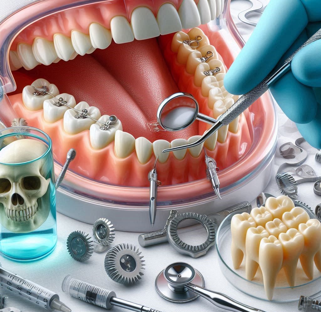
- Orthodontic Appliances:
- Braces: Traditional braces are often used to correct tooth misalignment and bite problems. They apply continuous pressure to move teeth into their correct positions.
- Functional Appliances: Devices like the Herbst appliance, Twin Block appliance, and headgear are used to influence the growth of the jaw in younger patients. These appliances encourage forward movement of the lower jaw or help correct other skeletal imbalances.
- Orthognathic Surgery: In more severe cases, where skeletal malocclusions cannot be corrected through orthodontic treatment alone, orthognathic surgery may be necessary. This surgery involves repositioning the jawbones to correct skeletal discrepancies and improve function and appearance.
- Surgical Timing: Typically performed after the growth phase, usually in late adolescence or early adulthood, to ensure that the facial bones have reached their final size and shape.
- Recovery and Post-Surgical Orthodontics: After surgery, orthodontic treatment is typically needed to perfect the alignment of the teeth.
The Role of the Orthodontist in Treating Supernumerary Teeth and Skeletal Malocclusions
Orthodontists are essential in diagnosing and managing dental conditions such as supernumerary teeth (extra teeth) and skeletal malocclusions (misalignment of the jaws). Their role is crucial in both early detection and treatment to avoid long-term complications, including tooth displacement, jaw dysfunction, and aesthetic concerns.
A. Diagnostic Tools
To accurately assess the condition of the teeth and jaws, orthodontists rely on a variety of diagnostic tools:
- Clinical Examination: This is the initial step in diagnosis, where the orthodontist performs a detailed visual and physical inspection of the patient’s teeth, gums, and jaw structure. The goal is to detect any obvious malocclusions or anomalies, such as extra teeth or misaligned jaws.
- Radiographs: X-rays are essential in orthodontics for evaluating the positioning of teeth, roots, and jawbones. Panoramic X-rays provide a broad view of the entire dental structure, while cephalometric X-rays allow for precise measurements of the head and jaw, helping to assess the alignment and growth of the skeletal components.
- 3D Imaging: Cone Beam Computed Tomography (CBCT) is a highly advanced diagnostic tool that provides detailed, three-dimensional images of the teeth, jawbones, and surrounding tissues. It allows orthodontists to plan treatments more accurately, especially in cases where teeth are impacted or abnormal bone growth is present.
B. Treatment Planning
Once the evaluation is complete, the orthodontist designs a customized treatment plan tailored to the patient’s specific needs. The treatment approach may include several strategies:
- Extraction: For cases involving supernumerary teeth or impacted teeth, the orthodontist may recommend surgical extraction to prevent complications like crowding or tooth impaction.
- Orthodontic Appliances: Braces, clear aligners, or other orthodontic devices are commonly used to address misalignments in the teeth and jaw, correcting malocclusions and improving bite function.
- Monitoring Jaw Growth: In growing patients, orthodontists will carefully monitor the development of the jaw. Timing is critical, as early intervention can help guide bone growth and potentially reduce the need for future surgical interventions.
By combining these diagnostic and treatment methods, orthodontists can provide effective care, improving both function and appearance for patients.
Conclusion
Orthodontics plays a pivotal role in treating complex conditions like supernumerary teeth and skeletal malocclusions. These conditions require early detection, precise diagnosis, and a multifaceted treatment approach that combines surgical and non-surgical methods. By improving both the function and aesthetics of the teeth and jaw, orthodontics helps enhance the patient’s quality of life. Early intervention remains key to ensuring the best possible outcomes, as the success of treatment depends on the timing and method of intervention. Through ongoing research and advancements in orthodontic techniques and technologies, the ability to treat these conditions continues to improve, offering patients greater options and outcomes.
SOURCES
Proffit, W. R., & Fields, H. W. (2018). Contemporary orthodontics (6th ed.). Elsevier.
Thilander, B., & Myers, M. (2016). The prevalence of supernumerary teeth in the general population and its associations with other dental anomalies. European Journal of Orthodontics, 38(3), 264-269.
Bishara, S. E. (2001). Impact of orthodontic treatment on dental and skeletal development. Seminars in Orthodontics, 7(3), 156-164.
Jiang, X., Zhao, X., Zhang, Y., Xie, Y., & Li, X. (2019). A clinical study on supernumerary teeth and associated conditions in children. Journal of Clinical Pediatric Dentistry, 43(2), 111-117.
Garcia, R. I., Santos, P. M., & Souza, L. G. (2020). Orthodontic management of supernumerary teeth and its implications. Brazilian Journal of Oral Sciences, 19, e2020.
Gundlach, K. K., Baum, U., Jäger, A., & Südkamp, N. (2010). Impact of early diagnosis and treatment of skeletal malocclusions on facial growth. International Journal of Oral and Maxillofacial Surgery, 39(12), 1155-1163.
Graber, T. M., Vanarsdall, R. L., Lynch, C. D., & Dawson, L. (2016). Orthodontics: Current principles and techniques (6th ed.). Elsevier.
Moyers, R. E. (2005). Handbook of orthodontics (5th ed.). McGraw-Hill.
Tweed, C. H. (1966). Clinical orthodontics. Saunders.
McNamara, J. A., & Behrents, R. G. (2004). The craniofacial complex and its relationship to orthodontics: A review. American Journal of Orthodontics and Dentofacial Orthopedics, 125(5), 624-634.
Miller, M. A. (2007). Supernumerary teeth: A review of the literature and treatment options. Dental Clinics of North America, 51(2), 415-430.
Woodhouse, M. R., Mills, D. B., & Griffiths, S. G. (2017). The orthodontic management of malocclusions and associated skeletal problems. Journal of Orthodontics and Craniofacial Research, 16(1), 27-34.
Boucher, C. O., & McKendrick, W. (2006). Treatment of skeletal malocclusions with orthognathic surgery. Journal of Oral and Maxillofacial Surgery, 64(6), 973-978.
Moyers, R. E., & Graber, T. M. (2008). Handbook of orthodontics (6th ed.). McGraw-Hill.
Bishara, S. E., & Marcos, G. R. (2014). Management of supernumerary teeth and their complications. Journal of Clinical Orthodontics, 48(9), 547-554.
Staley, R. N. (1996). The role of orthodontics in managing jaw deformities and malocclusions. American Journal of Orthodontics and Dentofacial Orthopedics, 110(1), 12-19.
Barton, G. M., & Brodie, A. G. (2012). Skeletal malocclusion and its correction in the adult. Journal of the American Dental Association, 143(8), 905-912.
Hägg, U., & Tarvonen, H. (2001). The effect of early orthodontic treatment on facial growth in patients with skeletal Class II malocclusions. European Journal of Orthodontics, 23(1), 67-72.
Pinho, T. S., Lima, G. R., & Teixeira, M. M. (2019). Current approaches to the treatment of Class III malocclusion. Orthodontic Perspectives, 22(3), 125-133.
Laskin, D. M., & Wheeler, T. (2010). An analysis of temporomandibular joint dysfunction associated with skeletal malocclusions. Journal of Oral Surgery, 68(1), 45-51.
HISTORY
Current Version
March 11, 2025
Written By:
SUMMIYAH MAHMOOD